Saturday, 23 October 2010
World's most powerful x-ray machine
After years of design and construction, the world's brightest X-ray machine has come to life at the SLAC National Accelerator Laboratory, in the hills near Stanford University. The mile-long machine produces a probing laser beam made of X-rays instead of visible light. Its laser bursts are so bright and so brief that researchers will use them as an ultrafast stop-motion camera to capture the minute details of things previously unseen, such as the arrangement of atoms in metals, semiconductors, ceramics, polymers and proteins.
Improved Medical Equipment for the Obese
Improved Medical Equipment for the Obese New X-ray Machine Handles Patients Up to 600 Pounds According to the Centers for Disease Control and Prevention, there has been a dramatic increase in obesity over the last two decades. With expanding waistlines also come increased health risks and more demand for bariatric surgery, gastric bypasses and lap bands.
Due to the lack of medical equipment designed to cater to their size, people who suffer from obesity often experience difficulty getting quality healthcare. In response to the increased size of patients, the medical industry is using innovation and technology to raise the standard of care.
A new X-ray machine, the Axiom Luminos TF, can handle people weighing up to 600 pounds. Described as an X-ray video camera, it allows doctors to see what's going on inside the body in real time. It also has a larger gap between the table top and the tower - which shoots a much wider picture.
With improvements like these, people undergoing surgery can be monitored in comfort within hours of the operation and in the following months; giving them the same level of care as the average sized person.
Tuesday, 12 October 2010
Medical Ventilators
Mechanical ventilation can be used to provide complete support, which means the ventilator is doing on the breathing for the patient. It can also be used to assist with breathing. This means the patient is still breathing on his own, but the ventilator is providing some assistance.

Before a person is placed on a medical ventilator, a breathing tube will be inserted into the airway, usually through the mouth. In some cases, a tracheotomy is performed instead. An incision is made in the trachea, and a tracheotomy tube is placed. This is usually done if long-term ventilator support is needed.
Ventilators can be set to different modes, which provide varied levels of support. A respiratory rate and a percentage of oxygen can be set. Mechanical ventilation is most often used in hospitals and can be complex.
The length of time a person stays on a medical ventilator depends on their condition. Patients who are only on a ventilator for surgery are usually extubated, which means the breathing tube and ventilator are removed. People with medical conditions that cause breathing problems may need to stay on a medical ventilator until the condition is treated and breathing improves.
Some individuals may need a mechanical ventilator for the rest of their lives. Conditions such as certain spinal cord injuries and severe brain damage may require continuous mechanical ventilation. Ventilators intended for home use are available.
When a patient is intubated, a sedative will be delivered through intravenous (IV) therapy. Sedatives are used to stop patients from fighting the ventilator which could cause damage to the lungs. Sedation may also lessen the pain and irritation of the throat or nasal passage associated with intubation.
Negative pressure is the oldest form of mechanical ventilation. The iron lung, a huge machine that stretches from the neck to the lower abdomen, was the first used in 1929 to treat breathing problems. The negative pressure used to stimulate breathing expands the abdomen which can cut off blood circulation to the lower body. Regular movement of the legs is required to stop blood from pooling in the extremities.
Iron lungs are rarely used today due to machine size and inability of the patient to move. Negative pressure, however can be a lifesaving form of mechanical ventilation. The Biphasic Cuirass is a modern form of the iron lung used in patients that cannot or should not be intubated. The cuirass machine works with the same negative pressure as the iron lung, but is much smaller and lighter.
Mechanical ventilation is meant to be a short term treatment. Injury to the airway, lungs, and pneumonia are risks associated with forced breathing. Patients are often weaned off artificial respiration slowly by using spontaneous breathing trials to judge whether the lungs will work properly without ventilation.
LASIK
LASIK is an acronym for LASer In-situ Keratomileusis, which simply means "to shape the cornea within using a laser." It corrects vision by reshaping the cornea (outer window of the eye) so that light rays focus more precisely on the retina, thereby reducing or eliminating refractive errors.

Using an instrument known as a microkeratome, a thin protective flap of corneal tissue is folded back.
The Excimer laser then removes a predetermined amount of tissue from the inner cornea to correct the individual's refractive error. The cornea is made flatter to treat nearsightedness, steeper to treat farsightedness and/or more spherical to correct astigmatism.
The corneal flap is placed back in its original position where it bonds without the need for stitches. LASIK can treat low to very severe refractive errors.
 The process:

Using an instrument known as a microkeratome, a thin protective flap of corneal tissue is folded back.
The Excimer laser then removes a predetermined amount of tissue from the inner cornea to correct the individual's refractive error. The cornea is made flatter to treat nearsightedness, steeper to treat farsightedness and/or more spherical to correct astigmatism.
The corneal flap is placed back in its original position where it bonds without the need for stitches. LASIK can treat low to very severe refractive errors.

- After your eye has been numbed with "eye drop" anesthesia, an instrument known as an eyelid speculum will be positioned to hold your eyelids open. You will remain awake and comfortable throughout the procedure.
- A small suction ring will be placed around the cornea and serves as a platform for the microkeratome.
- The microkeratome separates the surface layers of the cornea, and the corneal flap is folded back.
- You will be asked to look at a target light while the Excimer laser reshapes the corneal tissue. A clicking sound can be heard as each microscopic layer of tissue is vaporized. This process will last from seconds to minutes, depending on the amount of correction necessary.
- The corneal flap is then placed back into its original position and allowed to dry for a few minutes.
- You will be given additional eye drops, and your eye may be shielded for protection. Your vision will probably be a little blurry at first so have someone drive you home and relax for the rest of the day.
Monday, 11 October 2010
Medical Lasers History
Light Amplification by the Stimulated Emission of Radiation was originally described as a theoretical concept by Albert Einstein in 1917, but it was not until 1954 that the first "stimulated" emissions of microwave radiation (MASER) were generated by J.P. Gordon and C.H. Townes at Bell Laboratories.

Theoretical calculations for the construction of a visible light MASER, or LASER were published in 1958. The first LASER was built in 1960 by Dr. T.H. Maiman at Hughes Aircraft Company, using a synthetic ruby rod stimulated by high intensity flashlamps, which generated millisecond pulses of coherent 694nm Ruby Laser (red) light . Shortly afterwards, 1060nm (near-infrared) laser light was generated by stimulating glass rods doped with Neodymium (Nd:Glass Laser).
Within a year, pioneers such as Dr. Leon Goldman began research on the interaction of laser light on biologic systems, including early clinical studies on humans. Interest in medical applications was intense, but the difficulty controlling the power output and delivery of laser energy, and the relatively poor absorption of these red and infrared wavelengths led to inconsistent and disappointing results in early experiments. The exception was the application of the Ruby Laser in retinal surgery in the mid-60's. In 1964, the Argon Ion Laser was developed. This continuous wave 488nm (blue-green) gas laser was easy to control, and it's high absorption by hemoglobin made it well suited to retinal surgery, and clinical systems for treatment of retinal diseases were soon available.

In 1964, the Nd:YAG (Neodymium:Yttrium Aluminum Garnet) Laser and CO2 (Carbon Dioxide) Laser were developed at Bell Laboratories. The CO2 laser is a continuous wave gas laser, and emitted infrared light at 10600nm in an easily manipulated, focused beam that was well absorbed by water. Because soft tissue consists mostly of water, researchers found that a CO2 laser beam could cut tissue like a scalpel, but with minimal blood loss. The surgical uses of this laser were investigated extensively from 1967-1970 by pioneers such as Dr. Thomas Polanyi and Geza Jako, and in the early 70's, use of the CO2 laser in ENT and gynecologic surgery became well established, but was limited to academic and teaching hospitals.
Dye Lasers became available in 1969, and noble gas-halide, or Excimer Lasers in 1975. Since then, many other different laser systems have become available for industrial scientific, telecommunication, as well as medical use.
In the early 1980's, smaller but more powerful lasers became available, and were soon appearing in community hospitals and even physician's offices. Most of these systems were CO2 lasers used for cutting and vaporizing, and Argon lasers for opthalmic use. Nd:YAG and KTP laser systems were used by larger hospitals for the new field of laparoscopic surgery. These "second generation" lasers were all continous wave, or CW systems, which tend to cause non-selective heat injury, and proper use required a long "learning curve" and experienced laser surgeons.
The single most significant advance in the use of medical lasers was the concept of "pulsing" the laser beam, which allowed selective destruction of abnormal or undesired tissue, while leaving surrounding normal tissue undisturbed. The first lasers to fully exploit this principal of "selective thermolysis" were the Pulsed Dye Lasers introduced in the late 1980's for the treatment of port wine stains and strawberry marks in children, and shortly after, the first Q-switched lasers for the treatment of tattoos. Another major advance was the introduction of scanning devices in the early 1990s, enabling precision computerized control of laser beams. Scanned, pulsed lasers revolutionized the practice of plastic and cosmetic surgery by making safe, consistent laser resurfacing possible, as well as increasing public awareness of laser medicine and surgery.
Medical lasers have made it possible to treat conditions which previously were untreatable, or difficult to treat. Patients benefit by improved results and less cost. In the last few years, the main focus of research and development of medical lasers has been on Laser Hair Removal , the treatment of vascular lesions including Leg Veins, and vision correction. The thrust of current research is directed towards non-ablative laser resurfacing ("laser skin toning"), "no-touch" computerized vision correction, and improved photodynamic therapy for treatment of skin cancer and for hair removal

Within a year, pioneers such as Dr. Leon Goldman began research on the interaction of laser light on biologic systems, including early clinical studies on humans. Interest in medical applications was intense, but the difficulty controlling the power output and delivery of laser energy, and the relatively poor absorption of these red and infrared wavelengths led to inconsistent and disappointing results in early experiments. The exception was the application of the Ruby Laser in retinal surgery in the mid-60's. In 1964, the Argon Ion Laser was developed. This continuous wave 488nm (blue-green) gas laser was easy to control, and it's high absorption by hemoglobin made it well suited to retinal surgery, and clinical systems for treatment of retinal diseases were soon available.

Dye Lasers became available in 1969, and noble gas-halide, or Excimer Lasers in 1975. Since then, many other different laser systems have become available for industrial scientific, telecommunication, as well as medical use.
In the early 1980's, smaller but more powerful lasers became available, and were soon appearing in community hospitals and even physician's offices. Most of these systems were CO2 lasers used for cutting and vaporizing, and Argon lasers for opthalmic use. Nd:YAG and KTP laser systems were used by larger hospitals for the new field of laparoscopic surgery. These "second generation" lasers were all continous wave, or CW systems, which tend to cause non-selective heat injury, and proper use required a long "learning curve" and experienced laser surgeons.
The single most significant advance in the use of medical lasers was the concept of "pulsing" the laser beam, which allowed selective destruction of abnormal or undesired tissue, while leaving surrounding normal tissue undisturbed. The first lasers to fully exploit this principal of "selective thermolysis" were the Pulsed Dye Lasers introduced in the late 1980's for the treatment of port wine stains and strawberry marks in children, and shortly after, the first Q-switched lasers for the treatment of tattoos. Another major advance was the introduction of scanning devices in the early 1990s, enabling precision computerized control of laser beams. Scanned, pulsed lasers revolutionized the practice of plastic and cosmetic surgery by making safe, consistent laser resurfacing possible, as well as increasing public awareness of laser medicine and surgery.
Medical lasers have made it possible to treat conditions which previously were untreatable, or difficult to treat. Patients benefit by improved results and less cost. In the last few years, the main focus of research and development of medical lasers has been on Laser Hair Removal , the treatment of vascular lesions including Leg Veins, and vision correction. The thrust of current research is directed towards non-ablative laser resurfacing ("laser skin toning"), "no-touch" computerized vision correction, and improved photodynamic therapy for treatment of skin cancer and for hair removal
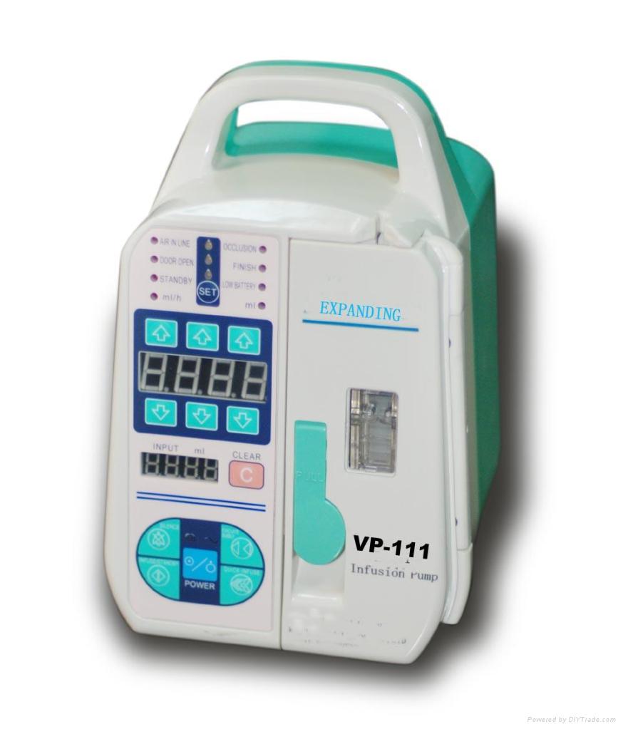
Infusion pump
An infusion pump infuses fluids, medication or nutrients into a patient's circulatory system. It is generally used intravenously, although subcutaneous, arterial and epidural infusions are occasionally used.
Infusion pumps can administer fluids in ways that would be impractically expensive or unreliable if performed manually by nursing staff. For example, they can administer as little as 0.1 mL per hour injections (too small for a drip), injections every minute, injections with repeated boluses requested by the patient, up to maximum number per hour (e.g. in patient-controlled analgesia), or fluids whose volumes vary by the time of day.
Because they can also produce quite high but controlled pressures, they can inject controlled amounts of fluids subcutaneously (beneath the skin), or epidurally (just within the surface of the central nervous system- a very popular local spinal anesthesia for childbirth).


Within these classes, some pumps are designed to be portable, others are designed to be used in a hospital, and there are special systems for charity and battlefield use.
Large-volume pumps usually use some form of peristaltic pump. Classically, they use computer-controlled rollers compressing a silicone-rubber tube through which the medicine flows. Another common form is a set of fingers that press on the tube in sequence.
Small-volume pumps usually use a computer-controlled motor turning a screw that pushes the plunger on a syringe.
The classic medical improvisation for an infusion pump is to place a blood pressure cuff around a bag of fluid. The battlefield equivalent is to place the bag under the patient. The pressure on the bag sets the infusion pressure. The pressure can actually be read-out at the cuff's indicator. The problem is that the flow varies dramatically with the patient's blood pressure (or weight), and the needed pressure varies with the administration route, making this quite risky for use by an untrained y less than 8 lbf/in² (55 kPa. Epidural and subcutaneous pressures are usually less than 18 lbf/in² (125 kPa).
Places that must provide the least-expensive care often use pressurized infusion systems. One common system has a purpose-designed plastic "pressure bottle" pressurized with a large disposable plastic syringe. A combined flow restrictor, air filter and drip chamber helps a nurse set the flow. The parts are reusable, mass-produced sterile plastic, and can be produced by the same machines that make plastic soft-drink bottles and caps. A pressure bottle, restrictor and chamber requires more nursing attention than electronically-controlled pumps. In the areas where these are used, nurses are often volunteers, or very inexpensive.
The restrictor and high pressure helps control the flow better than the improvised schemes because the high pressure through the small restrictor orifice reduces the variation of flow caused by patients' blood pressures.
An air filter is an essential safety device in a pressure infusor, to keep air out of the patients' veins: doctors estimate that 0.55 cm³ of air per kilogram of body weight is enough to kill (200–300 cm³ for adults) by filling the patient's heart. Small bubbles could cause harm in arteries, but in the veins they pass through the heart and leave in the patients' lungs. The air filter is just a membrane that passes gas but not fluid or pathogens. When a large air bubble reaches it, it bleeds off.
Some of the smallest infusion pumps use osmotic power. Basically, a bag of salt solution absorbs water through a membrane, swelling its volume. The bag presses medicine out. The rate is precisely controlled by the salt concentrations and pump volume. Osmotic pumps are usually recharged with a syringe.
Spring-powered clockwork infusion pumps have been developed, and are sometimes still used in veterinary work and for ambulatory small-volume pumps. They generally have one spring to power the infusion, and another for the alarm bell when the infusion completes.
Battlefields often have a need to perfuse large amounts of fluid quickly, with dramatically changing blood pressures and patient condition. Specialized infusion pumps have been designed for this purpose, although they have not been deployed.
Many infusion pumps are controlled by a small embedded system. They are carefully designed so that no single cause of failure can harm the patient. For example, most have batteries in case the wall-socket power fails. Additional hazards are uncontrolled flow causing an overdose, uncontrolled lack of flow, causing an underdose, reverse flow, which can siphon blood from a patient, and air in the line, which can starve a patient's tissues of oxygen if it floats to some part of a patient's body.
Infusion pumps can administer fluids in ways that would be impractically expensive or unreliable if performed manually by nursing staff. For example, they can administer as little as 0.1 mL per hour injections (too small for a drip), injections every minute, injections with repeated boluses requested by the patient, up to maximum number per hour (e.g. in patient-controlled analgesia), or fluids whose volumes vary by the time of day.
Because they can also produce quite high but controlled pressures, they can inject controlled amounts of fluids subcutaneously (beneath the skin), or epidurally (just within the surface of the central nervous system- a very popular local spinal anesthesia for childbirth).
Types of infusion
The user interface of pumps usually requests details on the type of infusion from the technician or nurse that sets them up:- Continuous infusion usually consists of small pulses of infusion, usually between 500 nanoliters and 10000 microliters, depending on the pump's design, with the rate of these pulses depending on the programmed infusion speed.
- Intermittent infusion has a "high" infusion rate, alternating with a low programmable infusion rate to keep the cannula open. The timings are programmable. This mode is often used to administer antibiotics, or other drugs that can irritate a blood vessel.
- Patient-controlled is infusion on-demand, usually with a preprogrammed ceiling to avoid intoxication. The rate is controlled by a pressure pad or button that can be activated by the patient. It is the method of choice for patient-controlled analgesia (PCA), in which repeated small doses of opioid analgesics are delivered, with the device coded to stop administration before a dose that may cause hazardous respiratory depression is reached.
- Total parenteral nutrition usually requires an infusion curve similar to normal mealtimes.

Types of pump
There are two basic classes of pumps. Large volume pumps can pump nutrient solutions large enough to feed a patient. Small-volume pumps infuse hormones, such as insulin, or other medicines, such as opiates.Within these classes, some pumps are designed to be portable, others are designed to be used in a hospital, and there are special systems for charity and battlefield use.
Large-volume pumps usually use some form of peristaltic pump. Classically, they use computer-controlled rollers compressing a silicone-rubber tube through which the medicine flows. Another common form is a set of fingers that press on the tube in sequence.
Small-volume pumps usually use a computer-controlled motor turning a screw that pushes the plunger on a syringe.
The classic medical improvisation for an infusion pump is to place a blood pressure cuff around a bag of fluid. The battlefield equivalent is to place the bag under the patient. The pressure on the bag sets the infusion pressure. The pressure can actually be read-out at the cuff's indicator. The problem is that the flow varies dramatically with the patient's blood pressure (or weight), and the needed pressure varies with the administration route, making this quite risky for use by an untrained y less than 8 lbf/in² (55 kPa. Epidural and subcutaneous pressures are usually less than 18 lbf/in² (125 kPa).
Places that must provide the least-expensive care often use pressurized infusion systems. One common system has a purpose-designed plastic "pressure bottle" pressurized with a large disposable plastic syringe. A combined flow restrictor, air filter and drip chamber helps a nurse set the flow. The parts are reusable, mass-produced sterile plastic, and can be produced by the same machines that make plastic soft-drink bottles and caps. A pressure bottle, restrictor and chamber requires more nursing attention than electronically-controlled pumps. In the areas where these are used, nurses are often volunteers, or very inexpensive.
The restrictor and high pressure helps control the flow better than the improvised schemes because the high pressure through the small restrictor orifice reduces the variation of flow caused by patients' blood pressures.
An air filter is an essential safety device in a pressure infusor, to keep air out of the patients' veins: doctors estimate that 0.55 cm³ of air per kilogram of body weight is enough to kill (200–300 cm³ for adults) by filling the patient's heart. Small bubbles could cause harm in arteries, but in the veins they pass through the heart and leave in the patients' lungs. The air filter is just a membrane that passes gas but not fluid or pathogens. When a large air bubble reaches it, it bleeds off.
Some of the smallest infusion pumps use osmotic power. Basically, a bag of salt solution absorbs water through a membrane, swelling its volume. The bag presses medicine out. The rate is precisely controlled by the salt concentrations and pump volume. Osmotic pumps are usually recharged with a syringe.
Spring-powered clockwork infusion pumps have been developed, and are sometimes still used in veterinary work and for ambulatory small-volume pumps. They generally have one spring to power the infusion, and another for the alarm bell when the infusion completes.
Battlefields often have a need to perfuse large amounts of fluid quickly, with dramatically changing blood pressures and patient condition. Specialized infusion pumps have been designed for this purpose, although they have not been deployed.
Many infusion pumps are controlled by a small embedded system. They are carefully designed so that no single cause of failure can harm the patient. For example, most have batteries in case the wall-socket power fails. Additional hazards are uncontrolled flow causing an overdose, uncontrolled lack of flow, causing an underdose, reverse flow, which can siphon blood from a patient, and air in the line, which can starve a patient's tissues of oxygen if it floats to some part of a patient's body.
Positron Emission Tomography (PET)
Positron emission tomography (PET) is a test that uses a special type of camera and a tracer (radioactive chemical) to look at organs in the body. The tracer usually is a substance (such as glucose) that can be used (metabolized) by cells in the body.

During the test, the tracer liquid is put into a vein (intravenous, or IV) in your arm. The tracer moves through your body, where much of it collects in the specific organ or tissue. The tracer gives off tiny positively charged particles (positrons). The camera records the positrons and turns the recording into pictures on a computer.
PET scan pictures do not show as much detail as computed tomography (CT) scans or magnetic resonance imaging (MRI) because the pictures show only the location of the tracer. The PET picture may be matched with those from a CT scan to get more detailed information about where the tracer is located.
A positron emission tomography (PET) scan is done to:

ref.: www.webmd.com

During the test, the tracer liquid is put into a vein (intravenous, or IV) in your arm. The tracer moves through your body, where much of it collects in the specific organ or tissue. The tracer gives off tiny positively charged particles (positrons). The camera records the positrons and turns the recording into pictures on a computer.
PET scan pictures do not show as much detail as computed tomography (CT) scans or magnetic resonance imaging (MRI) because the pictures show only the location of the tracer. The PET picture may be matched with those from a CT scan to get more detailed information about where the tracer is located.
A positron emission tomography (PET) scan is done to:
- Study the brain's blood flow and metabolic activity. A PET scan can help a doctor find nervous system problems, such as Alzheimer's disease, Parkinson's disease, multiple sclerosis, transient ischemic attack (TIA), amyotrophic lateral sclerosis (ALS), Huntington's disease, stroke, and schizophrenia.
- Find changes in the brain that may cause epilepsy.
- Evaluate some cancers, especially lymphoma or cancers of the head and neck, brain, lung, colon, or prostate. In its early stages, cancer may show up more clearly on a PET scan than on a CT scan or an MRI.
- See how advanced a cancer is and whether it has spread to another area of the body (metastasized). It is often necessary to do both CT and PET scans to evaluate cancer.
- Help a doctor choose the best treatment for cancer. PET scans may also be done to see whether surgery can be done to remove a tumor.
- Find poor blood flow to the heart, which may mean coronary artery disease.
- Find damaged heart tissue, especially after a heart attack.
- Help choose the best treatment, such as coronary artery bypass graft surgery, for a person with heart disease.

ref.: www.webmd.com
Saturday, 9 October 2010
Radiation Therapy
Radiation Therapy is a highly specialised area of the radiation sciences. It is a scientific and clinical profession dedicated to the management of patients with benign and malignant disease.
The ionising radiation in its commonest form for treatment is photons (x-rays). The radiation can be used on its own, or in combination with other cancer management strategies such a surgery or cytotoxic chemotherapy. This depends on many factors including the histological diagnosis, how advanced the disease is and the health of the patient.
The radiation can be delivered with one of two intensions radical and palliative. Radical intent is where high doses of radiation (depending on the histological diagnosis) are delivered with the intent to cure, and palliative intent is treatment in order to provide relief from cancer symptoms. One of the main considerations in managing cancer with radiation (as with any other treatment) is maintenance of quality of life for the patient.

The following steps constitute the radiation therapy process:
Integration of the above modalities in the localisation of the treatment ensures that even microscopic tumour cells and/or lymph nodes that are positively identified as having tumour present are included.
Radiation therapy planning utilises sophisticated computer systems to maximise tumour dose and minimise the dose to healthy surrounding tissues. A computer is used to generate a pictorial arrangement of the distribution of the radiation dose that the tumour and surrounding organs will receive.
This is imperative because organs such as the spinal cord or lungs can only tolerate minimal doses of radiation before irreparable damage occurs.
For any one particular treatment site the radiation beam can be directed towards the patient from a number of angles (fields) in order to reduce the dose to radiosensitive organs and ensure a high dose region around the tumour volume.
Radiation can be administered externally with machines that work at either Kilovoltage energies (used for treating superficial tumours) or Megavoltage energies (Linear Accelerators which are used to treat deep seated tumours).
Internal radioactive sources are also used to treat tissues and organs such as the tongue, cervix or prostate gland.

Daily treatment has to be both accurate and reproducible. This means that the patient needs to be immobilised in exactly the same position every day. Precise measurements are used to align the radiation beam with the specific area of the body being treated. The treatment area itself can be verified on the treatment machine before, during or after the daily treatment is delivered. These images can then be matched with those from the original planning procedure using sophisticated computer equipment.
Prior to any patients being treated all equipment must go through rigorous quality assurance procedures in order to ensure it is operating safely.
The technology within the field of radiation therapy is constantly developing with the view of achieving optimal conformation of the radiation beam to the tumour volume. Utilisation of Intensity Modulated Radiation Therapy (IMRT) is the closest the technology has got to achieving this.
ref.: http://www.med.monash.edu.au/radiography
The ionising radiation in its commonest form for treatment is photons (x-rays). The radiation can be used on its own, or in combination with other cancer management strategies such a surgery or cytotoxic chemotherapy. This depends on many factors including the histological diagnosis, how advanced the disease is and the health of the patient.
The radiation can be delivered with one of two intensions radical and palliative. Radical intent is where high doses of radiation (depending on the histological diagnosis) are delivered with the intent to cure, and palliative intent is treatment in order to provide relief from cancer symptoms. One of the main considerations in managing cancer with radiation (as with any other treatment) is maintenance of quality of life for the patient.

The following steps constitute the radiation therapy process:
Localisation
This is the initial step in the radiotherapy process. At this point the exact position of the tumour is located by utilising diagnostic image acquisition techniques such as plain radiographs, CT, MRI and PET. Simulation of the treatment area may also occur where the treatment ‘set up’ is reproduced and radiographic images of this are acquired and recorded. These images are then interpreted and used to configure individual treatment plans for each patient and also for comparison during treatment itself.Integration of the above modalities in the localisation of the treatment ensures that even microscopic tumour cells and/or lymph nodes that are positively identified as having tumour present are included.
Planning
The tumour/site of original tumour and an area of tissue surrounding it are treated to the highest possible dose. This combined site of tumour and normal cells is known as the Tumour Volume.Radiation therapy planning utilises sophisticated computer systems to maximise tumour dose and minimise the dose to healthy surrounding tissues. A computer is used to generate a pictorial arrangement of the distribution of the radiation dose that the tumour and surrounding organs will receive.
This is imperative because organs such as the spinal cord or lungs can only tolerate minimal doses of radiation before irreparable damage occurs.
For any one particular treatment site the radiation beam can be directed towards the patient from a number of angles (fields) in order to reduce the dose to radiosensitive organs and ensure a high dose region around the tumour volume.
Treatment
There are a variety of radiation modalities available for treatment. The choice of radiation/particle type (photon, electron, proton, neutron, beta and gamma) and energy depends on a number of factors such as how ‘deep seated’ the tumour is and the nearby radiosensitive structures.Radiation can be administered externally with machines that work at either Kilovoltage energies (used for treating superficial tumours) or Megavoltage energies (Linear Accelerators which are used to treat deep seated tumours).
Internal radioactive sources are also used to treat tissues and organs such as the tongue, cervix or prostate gland.

Daily treatment has to be both accurate and reproducible. This means that the patient needs to be immobilised in exactly the same position every day. Precise measurements are used to align the radiation beam with the specific area of the body being treated. The treatment area itself can be verified on the treatment machine before, during or after the daily treatment is delivered. These images can then be matched with those from the original planning procedure using sophisticated computer equipment.
Prior to any patients being treated all equipment must go through rigorous quality assurance procedures in order to ensure it is operating safely.
The technology within the field of radiation therapy is constantly developing with the view of achieving optimal conformation of the radiation beam to the tumour volume. Utilisation of Intensity Modulated Radiation Therapy (IMRT) is the closest the technology has got to achieving this.
ref.: http://www.med.monash.edu.au/radiography
DEXA
DEXA is a dual energy X-ray absorptiometry imaging technique widely used for non-invasive assessment of bone mineral content. This has a direct impact on the management and treatment on the condition of osteoporosis.

Osteoporosis, within our community, continues to debilitate those with the condition and presents a large problem for those with an increased risk of fracture. This imaging system allows for early detection of the condition and acts as a baseline study for future following preventative management and treatments.
ref.: http://www.med.monash.edu.au/radiography

ref.: http://www.med.monash.edu.au/radiography
Digital Vascular Imaging
This is an imaging modality that utilises the technology of digital fluoroscopy and additional equipment and computer systems to image the blood vessels (arteries and veins) of the human body.

The images produced serve a diagnostic purpose; that is, diagnosing a pathology or condition. Also, treatment or therapeutic cases can be performed such as stenting (inserting a device into a blood vessel in order to keep it open and allow blood to flow through) or infusion of thrombolytic agents (administering a medication such as Urokinase to help breakdown a recently formed thromus or blood clot).
The procedures are performed under sterile conditions and require that the patient be fasted (no food prior to procedure) and the radiologist or cardiologist (the specialist medical practitioner who performs the invasive procedure and subsequently interprets the images) be "gowned" as in an operating theatre. Specialised DVI suites are used to image the blood vessels supplying blood to the heart itself.
This modality requires that the radiographer is part of a team approach working closely with other health professionals such as radiologists, cardiologists, nurses and cardiac technicians
ref.: http://www.med.monash.edu.au/radiography
Friday, 8 October 2010
Magnetic Resonance Imaging or MRI
This is an advanced and specialised field of radiography and medical imaging. The equipment used is very precise, sensitive and at the forefront of clinical technology. MRI is not only used in the clinical setting, it is increasingly playing a role in many research investigations.

Magnetic Resonance Imaging (MRI) utilises the principle of Nuclear Magnetic Resonance (NMR) as a foundation to produce highly detailed images of the human body.
The patient is firstly placed within a magnetic field created by a powerful magnet. The hydrogen atoms in the human body (we are made up of H2O) then align themselves with a North and South orientation within this magnetic field - that is, they behave like tiny bar magnets. Using a transmitting device, the radiographer transmits a radiofrequency (RF) pulse. This causes the hydrogen atoms to alter the direction of their orientation.
The transmitting RF pulse is switched off and the hydrogen atoms begin to return to the alignment they acquired when they were first placed in the magnetic field. As this re-alignment occurs they emit an RF signal which is detected by a receiving device or antenna.
From this received signal, sophisticated electronic and computer equipment is used to determine the intensity of this signal and the exact location from where this signal originated.
From this viewpoint, the computer performs advanced image reconstruction calculations and produces an image that can be viewed, hard and/or soft copied and interpreted for any diagnosis. The images can be acquired in a variety of planes, commonly, sagittal, axial and coronal (also oblique planes can be performed) - while the patient is lying still in the same position.
ref.: http://www.med.monash.edu.au/radiography
Mammography
Mammography uses dedicated, low-dose X-ray equipment, to obtain images of the breast to assist in the diagnosis of breast cancer and other breast diseases.

This is available to women through the Breastscreen Program and in Diagnostic Imaging facilities in both Public and Private Imaging Departments. Students in the third year of the Medical Imaging and Radiation Sciences course are given an understanding of the imaging process, the anatomy and pathology of the breast and an insight into the highly specialized communication and interpersonal skills required when dealing with both male and female patients and the diagnosis of breast disease.

Ultrasound
Ultrasound imaging is another of the many 'modalities' that is encountered in the imaging department. Its distinctive feature is that it uses high frequency ultrasound to construct an image rather than the traditional x-ray. This means that it is a safe, non-invasive means of creating cross sectional images of the human body. It is also a relatively cost-effective means of imaging.


Ultrasound however is used with great diversity beyond obstetrics. Vascular ultrasound, for instance, allows us to see the blood flow in real-time thus making it possible to discern stenoses in the arteries, or thrombosis of the veins. Musculoskeletal ultrasound allows us to image tiny tendons and nerves for degeneration or tears. Ultrasound is used in abdominal, gynaecological and paediatric assessment. The technology is enabling us to see the movement of organs, see their structure in 3D, and image their microvasculature.
Ultrasound imaging is performed either by a medical physician or a sonographer. The sonographer gains their accreditation through the Australian Sonographer Accreditation Registry (ASAR) after obtaining a two-year part time, Graduate Diploma in Medical Ultrasound. This is available through our department. Because of the emphasis upon sonography in the Bachelor of Radiography and Medical Imaging graduates with good grades in the sonographic units are eligible for exemption from several of the level one units in the Graduate Diploma. Sonographers work in multi-modality imaging centres in hospitals and private practices. They also work with specialist physicians such as obstetricians and vascular surgeons. Sonographers are involved in diagnosis and imaging.
ref.: http://www.med.monash.edu.au/radiography

Ultrasound is familiar to us all because of its role in obstetrics. Nearly all pregnant women, at some stage, experience the delight of seeing their developing fetus with this technology. While this is going on, they also experience an important medical test which will assist their management.

Ultrasound imaging is performed either by a medical physician or a sonographer. The sonographer gains their accreditation through the Australian Sonographer Accreditation Registry (ASAR) after obtaining a two-year part time, Graduate Diploma in Medical Ultrasound. This is available through our department. Because of the emphasis upon sonography in the Bachelor of Radiography and Medical Imaging graduates with good grades in the sonographic units are eligible for exemption from several of the level one units in the Graduate Diploma. Sonographers work in multi-modality imaging centres in hospitals and private practices. They also work with specialist physicians such as obstetricians and vascular surgeons. Sonographers are involved in diagnosis and imaging.
ref.: http://www.med.monash.edu.au/radiography
Wednesday, 6 October 2010
Computed Tomography
Computed tomography (CT) is an integral component of the general radiography department. Unlike conventional radiography, in CT the patient lies on a couch that moves through into the imaging gantry housing the x-ray tube and an array of specially designed "detectors". 
Depending upon the system the gantry rotates for either one revolution around the patient or continuously in order for the detector array to record the intensity of the remnant x-ray beam. These recordings are then computer processed to produce images never before thought possible. The familiar radiograph lacks a third dimension; it can only show us a two-dimensional view of the human body. CT on the other hand reconstructs images in a variety of body planes the most usual being the axial or cross sectional plane. The image created displays CT numbers which mainly reflect the physical properties of the tissues being investigated. Because of the large range of the CT number scale and the fact that the image is digital, it is possible to manipulate the display to show the underlying soft tissues with enhanced contrast as well as the bony structures.

Technological innovation has been a continuous feature of CT since its invention in 1971. Scanners today are capable of gathering even more data about the body structure in a time span that is measured in seconds thereby enhancing its clinical usefulness. CT innovation has meant radiographers today need to be able to recognize and evaluate anatomical structures in a variety of body planes.
ref.: http://www.med.monash.edu.au/radiography


ref.: http://www.med.monash.edu.au/radiography
Fluoroscopy
Traditionally fluoroscopy is an imaging method that uses x-rays and closed circuit television to produce "real time" images of the body.

Typically in clinical radiology departments fluoroscopy is used to image the digestive tract and the hepato-biliary system and genito-urinary system. In these cases radiographic contrast agents must be introduced into the patient in order to visualize organs that are normally only seen as shadows on a plain radiograph of the abdomen.

Increasingly tradition fluoroscopy is being replaced with digital systems that enhance its diagnostic capacity whilst reducing overall exposure levels. The mobile versions of the fluoroscopic system are used extensively in the operating theatre to assist surgeons to evaluate a wide variety of operative procedures.


ref.: http://www.med.monash.edu.au/radiography/
The Familiar X-Ray
The creation of the familiar plain radiograph begins with the radiographer receiving a request form for a radiographic examination of a particular part of a patient's body.

The next phase involves the radiographer assessing the patient prior to selecting the most appropriate imaging equipment and positioning methods for the projections that will best answer the clinical query. Essentially the radiographic procedure involves the selection of exposure factors and the accurate positioning of the patient's body in relation to the x-ray tube and the imaging device.

Today the imaging device will either be a conventional x-ray cassette and x-ray film which will be developed photographically or a digital plate which will be computer processed. Prior to sending the radiographs or images on for reporting by the radiologist, radiographers must evaluate their radiographs or images in terms of image quality, radiographic positioning and the clinical question. This means radiographers need a high level of knowledge about the science of image formation, radiographic anatomy and pathophysiology.

ref.: http://www.med.monash.edu.au/radiography
Tuesday, 5 October 2010
Definition Of Medical Imaging
Medical imaging is the technique and process used to create images of the human body (or parts and function thereof) for clinical purposes (medical procedures seeking to reveal, diagnose or examine disease) or medical science (including the study of normal anatomy and physiology).

Although imaging of removed organs and tissues can be performed for medical reasons, such procedures are not usually referred to as medical imaging, but rather are a part of pathology.
As a discipline and in its widest sense, it is part of biological imaging and incorporates radiology (in the wider sense), nuclear medicine, investigative radiological sciences, endoscopy, (medical) thermography, medical photography and microscopy (e.g. for human pathological investigations).
Measurement and recording techniques which are not primarily designed to produce images, such as electroencephalography (EEG), magnetoencephalography (MEG), Electrocardiography (EKG) and others, but which produce data susceptible to be represented as maps (i.e. containing positional information), can be seen as forms of medical imaging.
ref.: http://en.wikipedia.org
ref.: http://en.wikipedia.org
Definition of medical equipment
medical equipment is designed to aid in the diagnosis, monitoring or treatment of medical conditions. These devices are usually designed with rigorous safety standards.

There are several basic types:
- Diagnostic equipment includes medical imaging machines, used to aid in diagnosis. Examples are ultrasound and MRI machines, PET and CT scanners, and x-ray machines.
- Therapeutic equipment includes infusion pumps, medical lasers and LASIK surgical machines.
- Life support equipment is used to maintain a patient's bodily function. This includes medical ventilators, anaesthetic machines, heart-lung machines, ECMO, and dialysis machines.
- Medical monitors allow medical staff to measure a patient's medical state. Monitors may measure patient vital signs and other parameters including ECG, EEG, blood pressure, and dissolved gases in the blood.
- Medical laboratory equipment automates or helps analyze blood, urine and genes.
- Diagnostic Medical Equipment may also be used in the home for certain purposes, e.g. for the control of diabetes mellitus.
A biomedical equipment technician (BMET) is a vital component of the healthcare delivery system. Employed primarily by hospitals, BMETs are the people responsible for maintaining a facility's medical equipment.
ref.: http://en.wikipedia.org
Subscribe to:
Comments (Atom)